SARS-CoV-2 variants continue to be a topic of great concern. As weeks go by, it is evident that the Omicron family of viruses continue to grow in complexity. Figures one and two show the extent of variation from the beginning of the pandemic up to the present.

Figure 1 roughly illustrates the complexity of SARS-CoV-2 variants, with each strain labeled by its PANGO lineage designation. The vertical axis illustrates the number of people infected, and the horizontal axis displays the rate of spread of that particular variant in the population. Both axes are on a logarithmic scale. The relative growth rate is described as how many additional people are infected by one infected person, for instance, BA.2 exceeding 12. Reading the figure from left to right, the original set of variants before Delta in late 2020 and early 2021 is shown as a cloud of variants, distinguished by unique mutations around the common blueprint D614G Triad variant. Figure 2 illustrates the emergence of new variants by date and relative growth rate.
As we move from left to right across, we note that the rate of spread in populations increases. Notably, this occurs in many populations that have been either heavily infected, highly vaccinated, or both. The second cloud of variants comes from the Delta variant. These variants were more pathogenic and spread through the population more rapidly than the previous isolates.
There is then a gap as the emergence of new variants slowed. Over the past few months, the Delta cloud was displaced by two distinct families. The earlier was the BA.1 family of variants, followed by the current BA.2 family. Within the BA.2 family, many different variants have been identified, including some that fuel more infections than others, for example, the BA.2.12.1 variant spreading widely in the Northeast United States.

Outside of the United States, two new variants are gaining attention from the World Health Organization as they grow in their sequence and infection count: BA.4 ad BA.5, which are offshoots of the BA.2 family.
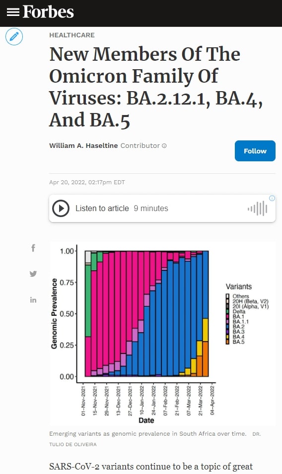
As the thumbnail of this article makes clear, BA.4 and BA.5 are growing rapidly to dominate the South African population. We note that just because instances of BA.4 and BA.5 are increasing as a relative proportion. That does not mean that the total number of cases is increasing in South Africa. Case rates are low and stable at the time of writing.
The Omicron subvariants BA.4 and BA.5 share most mutations with the original BA.2 Omicron variant, but each has its distinct mutations from BA.2 and one another. Here we examine these two subvariants and the new modifications they possess.
For reference, the BA.2 Spike protein contains 28 amino acid mutations compared to the original Wuhan strain. Fifteen of these are concentrated in the receptor-binding domain. In addition to the mutations in the receptor-binding domain, there are also a number of mutations in the N-terminal domain and the membrane-associated S2 region in the Spike.
The BA.4 and BA.5 subvariants share an identical Spike protein with four modifications relative to BA.2.
Receptor-Binding And N-Terminal Domain Mutations
The first of these four is a reversion of a mutation back to its original amino acid. The mutation of glutamine to arginine at position 493 (Q493R) in BA.2 returns to glutamine in BA.4 and BA.5. There are studies indicating that Q493R is an escape mutation for some monoclonal antibody treatments, such as bamlanivimab and etesevimab. A reversion to Q493 may suggest that BA.4 and BA.5 are more susceptible to neutralization by monoclonal antibodies, though further investigation would be needed.

The next mutation, leucine to arginine at position 452 (L452R), is common in existing variants. The mutation induces a change in charge from positive to neutral. The mutation is cited for contributing to immune escape from antibody binding and may also contribute to stronger cell attachment to the virus, leading to increased transmissibility and pathogenicity.
The least common of the three amino acid changes is phenylalanine to valine at position 486 (F486V). This mutation does not induce a polar or charge shift. Dr. Jesse Bloom suggests that F486V could “lead to more antibody escape from serum elicited by current vaccines / early infections,” though further research to confirm this hypothesis would be useful.
There is also a deletion of positions 69 and 70 in the N-terminal domain. This is a common mutation found in natural variants and likely knocks out an antibody binding site, meaning this specific mutation could impact overall immune evasion.
The rate at which the virus spreads through a population that has a large percentage of Covid-19 recoverees is determined both by the number of people who have been previously infected as well as the number of those vaccinated. A new variant must have the viral fitness to overcome immune defenses developed during previous infections and vaccinations to spread in such a population. One way of understanding escape from a waning immunity is to look at the sensitivity of variants to monoclonal antibodies based on certain escape mutations.
Despite the mutation at position 493 reverting to its original state, the inclusion of the N-terminal domain deletion, F486V, and L452R may yield a more immune evasive virus overall. Both receptor-binding domain mutations were cited by Dr. Tulio de Oliveira as being associated with escape neutralization from all classes of monoclonal antibodies.

The BA.4 and BA.5 subvariants also come with a few modifications to the genomic profile of the virus outside the Spike protein.

Nonstructural, Structural, and Accessory Protein Mutations
Both BA.4 and BA.5 revert two mutations back to their wild type: NSP4 L438 and Orf6 D61. The exact functions of these mutations are unknown, though we can speculate based on the function of their resident protein. NSP4 is involved in forming the double-membrane vesicle, which is involved in replication. Orf6 inhibits the innate immune response, downregulating various signals, proteins, and enzymes that impede virus replication.
BA.4 adds two mutations closer to the 3’ end of the genome: leucine to phenylalanine at position 11 (L11F) in Orf7b and proline to serine at position 151 (P151S) in the N protein. Both Orf7b and N are involved in the evasion of innate immunity, and these mutations may contribute to evasion efficiency. The N protein mutations may also affect the packaging and stability of the virus particle.
BA.5 adds aspartic acid to asparagine at position 3 (D3N) in the M protein. The Membrane protein surrounds the virus particle and is involved in viral entry, and this mutation may increase functional efficiency. The M protein also plays a role in the suppression of innate immunity. The D3N mutation is relatively uncommon, but D3G is found in some Omicron lineages.
These viruses should not be underestimated. For every person infected with the Wuhan strain, these viruses may infect roughly 12. Previous mitigation methods have spared China the level of infections rampant elsewhere. However, mild increases in cases in comparison seem to show it is more difficult to contain the spread of the Omicron variants.
Based on observations thus far, I doubt that SARS-CoV-2 has reached the upper limits of its rate of spread in either vaccinated or unvaccinated populations. I think new variants may emerge with substantially increased virulence. We should remember that a closely related virus to SARS-CoV-2—MERS-CoV—kills roughly 50% of those it infects.
The story of the omicron family of variants is still emerging and one that the world is watching closely. It is unfortunate that at a time when surveillance of infection rates and strain identification is crucial, those efforts are declining rapidly worldwide. This strikes as a grave mistake, as surveillance is the best early warning signal for new and perhaps more dangerous variants.


